
Uploads by Yp305
This special page shows all uploaded files.
| Date | Name | Thumbnail | Size | Description | Versions |
|---|---|---|---|---|---|
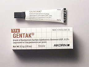
| 16:46, 15 November 2006 | Image8.JPG (file) |  |
33 KB | product of gentamysin | 1 |
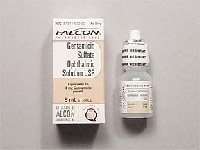
| 16:43, 15 November 2006 | Image7.JPG (file) |  |
14 KB | gentamysin product | 1 |
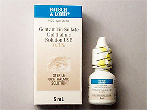
| 16:40, 15 November 2006 | Image6.JPG (file) |  |
34 KB | gentamysin eye drops | 1 |
| 16:30, 15 November 2006 | Image5.jpg (file) |  |
6 KB | structure of gentamysin action | 1 |
| 16:30, 15 November 2006 | Image4.jpg (file) |  |
8 KB | structure of gentamysin action | 1 |
| 16:26, 15 November 2006 | Image3.jpg (file) |  |
8 KB | structure of gentamysin action | 1 |
| 16:22, 15 November 2006 | Image2.jpg (file) |  |
6 KB | structure of gentamysin action | 1 |
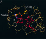
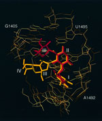
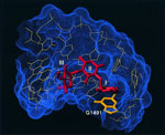
| 16:17, 15 November 2006 | Image1.jpg (file) |  |
8 KB | Binding pocket of gentamicin in the A-site RNA. The Connolly surface of the RNA is represented by blue dots and the gentamicin C1a is red. The view is from the major groove of the RNA. The three rings of gentamicin C1a are numbered as in Figure 1. The bas | 1 |
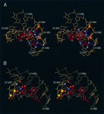
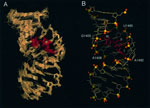
| 16:10, 15 November 2006 | 7591334f5a.jpg (file) |  |
68 KB | (A) Best-fit superposition of 38 final simulated annealing structures of the A-site RNA−gentamicin C1a complex, viewed from the major groove side of the RNA. The heavy atoms have been superimposed. The RNA is shown in beige and gentamicin C1a is red. Th | 1 |
| 15:26, 20 October 2006 | Gentamycin.cdx (file) | 2.43 MB | gentamycin in 3D form | 1 |